제품특징
Western Blotting Detection
Amersham ECL Select (Low picogram, <5pg)
Amersham ECL range 중 가장 감도가 높은 시약입니다. 단백질 발현의 작은 변화도 검출 가능하며, Low picograms(<5pg) 수준의 단백질까지 검출할 수 있습니다.
Amersham ECL Prime (Mid picogram, 10-100pg)
고감도 시약으로서, 시간에 따른 단백질 수준의 변화를 정확히 검출할 수 있도록 24시간 이상의 signal duration을 가집니다.
Amersham ECL (Mid picogram, 20-100pg)
Standard ECL 시약으로서, Mid picograms(20-100pg)수준의 단백질 검출이 가능합니다.
제품스펙
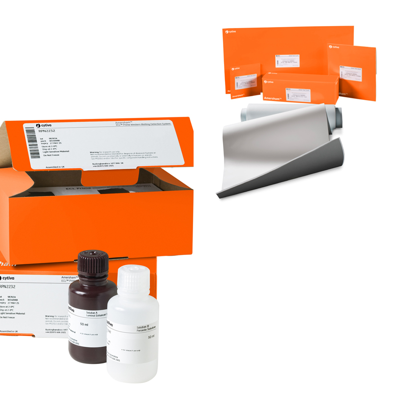
Amersham™ ECL™ Wester Blotting Detection Reagents
| 품명 |
규격 |
품번 |
| Amersham™ ECL Select™ |
100 ml |
RPN2235 |
| Amersham™ ECL™ Prime |
100 ml |
RPN2232 |
| Amersham™ ECL™ |
500 ml |
RPN2106 |
| 250 ml |
RPN2209 |
| 125 ml |
RPN2109 |
| Amersham™ ECL™ start |
400 ml |
RPN3244 |
| 200 ml |
RPN3243 |
관련 제품
| 구분 |
품명 |
규격 |
품번 |
| Membrane |
Amersham™ Hybond™ P PVDF 0.2 μm |
260 mm x 4 m |
10600021 |
| Amersham™ Hybond™ P PVDF 0.45 μm |
300 mm x 4 m |
10600023 |
| Amersham™ Protran™ NC 0.2 μm |
300 mm x 4 m |
10600001 |
| Amersham™ Protran™ NC 0.4 μm |
300 mm x 4 m |
10600002 |
| Marker |
Amersham™ ECL DualVue™ WB Markers |
25 gel loadings |
RPN810 |
| Amersham™ ECL™ Full-Range Rainbow Markers |
250 μl |
RPN800E |
| HRP conjugated secondary antibodies |
Amersham™ ECL™ Mouse IgG HRP-Linked Whole Antibody |
1 ml |
NA931-1ML |
| Amersham™ ECL™ Rabbit IgG HRP-Linked Whole Antibody |
1 ml |
NA934-1ML |
주요사항
Benchmarking data
Amersham ECL Select
Figure 8 shows a side-by-side comparison of
Amersham ECL Select with other high-sensitive
chemiluminescence detection reagents.Samples: Two-fold dilution series of HeLa cell lysate starting at 2.5 μg
Primary antibody dilution: 1:10 000
Secondary antibody dilution: 1:100 000
Detection: Amersham Imager 600, 3 min exposure |
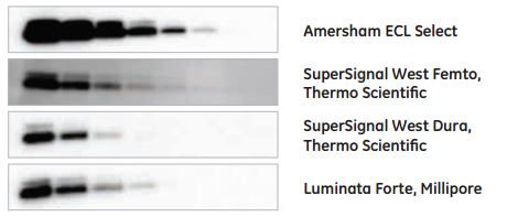
Fig 8. Amersham ECL Select shows brighter bands, lower background,
and higher limit of detection compared with competitor reagents in
Western blotting detection of ERK 1/2.
|
Amersham ECL Prime
Figure 6 shows a side-by-side comparison of
Amersham ECL Prime with other high-sensitive
chemiluminescence detection reagents.Samples: Two-fold dilution series of HeLa cell lysate starting at 10 μg
Primary antibody dilution: 1:5000
Secondary antibody dilution: 1:30 000
Detection: Amersham Imager 600, 75 s exposure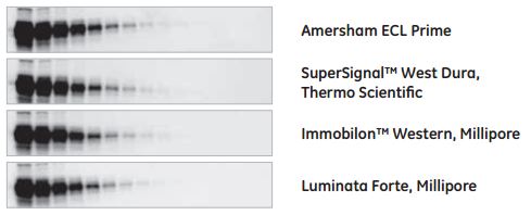 Fig 6. Amersham ECL Prime exhibits similar sensitivity as the competitor Fig 6. Amersham ECL Prime exhibits similar sensitivity as the competitor
reagents in Western blotting detection of ERK 1/2. The Millipore reagents
were slightly less sensitivity and also reached saturation at the highest
concentrations (10 to 5 μg), decreasing the dynamic range of these reagents. |
Figure 7 shows Amersham ECL Prime in comparison
with Clarity™ Western ECL Substrate from Bio-Rad.Samples: Two-fold dilution series of HeLa cell lysate starting at 2.5 μg
Primary antibody dilution: 1:3000
Secondary antibody dilution: 1:30 000
Detection: Amersham Imager 600, 3 min exposure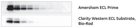 Fig 7. Amersham ECL Prime exhibits higher sensitivity and signal intensity Fig 7. Amersham ECL Prime exhibits higher sensitivity and signal intensity
compared with Clarity Western ECL Substrate in Western blotting
detection of ERK 1/2. |
Amersham ECL
Figure 5 shows a side-by-side comparison of
Amersham ECL reagent with other chemiluminescence
detection reagents in the same sensitivity range.Samples: Two-fold dilution series of HeLa cell lysate starting at 5 μg
Primary antibody dilution: 1:1000
Secondary antibody dilution: 1:10 000
Detection: Amersham Imager 600, 1 min exposure |
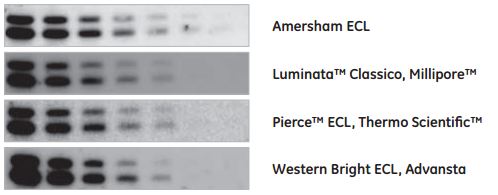
Fig 5. Amersham ECL reagent exhibits similar sensitivity as the competitor
reagents in Western blotting detection of ERK 1/2.
|

