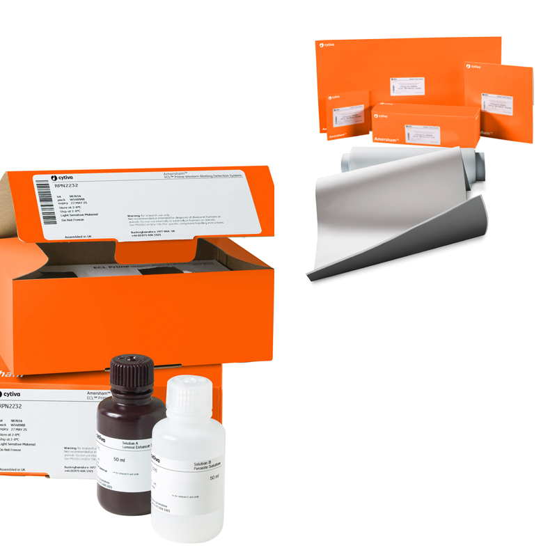
제품특징
Western Blotting Detection
Amersham ECL Select (Low picogram, <5pg)
Amersham ECL range 중 가장 감도가 높은 시약입니다. 단백질 발현의 작은 변화도 검출 가능하며, Low picograms(<5pg) 수준의 단백질까지 검출할 수 있습니다.
Amersham ECL Prime (Mid picogram, 10-100pg)
고감도 시약으로서, 시간에 따른 단백질 수준의 변화를 정확히 검출할 수 있도록 24시간 이상의 signal duration을 가집니다.
Amersham ECL (Mid picogram, 20-100pg)
Standard ECL 시약으로서, Mid picograms(20-100pg)수준의 단백질 검출이 가능합니다.
제품스펙
Amersham™ ECL™ Wester Blotting Detection Reagents
| 품명 |
규격 |
품번 |
| Amersham™ ECL Select™ |
100 ml |
RPN2235 |
| Amersham™ ECL™ Prime |
100 ml |
RPN2232 |
| Amersham™ ECL™ |
500 ml |
RPN2106 |
| 250 ml |
RPN2209 |
| 125 ml |
RPN2109 |
| Amersham™ ECL™ start |
400 ml |
RPN3244 |
| 200 ml |
RPN3243 |
관련 제품
| 구분 |
품명 |
규격 |
품번 |
| Membrane |
Amersham™ Hybond™ P PVDF 0.2 μm |
260 mm x 4 m |
10600021 |
| Amersham™ Hybond™ P PVDF 0.45 μm |
300 mm x 4 m |
10600023 |
| Amersham™ Protran™ NC 0.2 μm |
300 mm x 4 m |
10600001 |
| Amersham™ Protran™ NC 0.4 μm |
300 mm x 4 m |
10600002 |
| Marker |
Amersham™ ECL DualVue™ WB Markers |
25 gel loadings |
RPN810 |
| Amersham™ ECL™ Full-Range Rainbow Markers |
250 μl |
RPN800E |
| HRP conjugated secondary antibodies |
Amersham™ ECL™ Mouse IgG HRP-Linked Whole Antibody |
1 ml |
NA931-1ML |
| Amersham™ ECL™ Rabbit IgG HRP-Linked Whole Antibody |
1 ml |
NA934-1ML |
주요사항
Benchmarking data
Amersham ECL Select
Figure 8 shows a side-by-side comparison of
Amersham ECL Select with other high-sensitive
chemiluminescence detection reagents.Samples: Two-fold dilution series of HeLa cell lysate starting at 2.5 μg
Primary antibody dilution: 1:10 000
Secondary antibody dilution: 1:100 000
Detection: Amersham Imager 600, 3 min exposure |
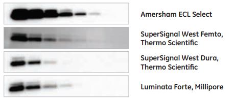
Fig 8. Amersham ECL Select shows brighter bands, lower background,
and higher limit of detection compared with competitor reagents in
Western blotting detection of ERK 1/2.
|
Amersham ECL Prime
Figure 6 shows a side-by-side comparison of
Amersham ECL Prime with other high-sensitive
chemiluminescence detection reagents.Samples: Two-fold dilution series of HeLa cell lysate starting at 10 μg
Primary antibody dilution: 1:5000
Secondary antibody dilution: 1:30 000
Detection: Amersham Imager 600, 75 s exposure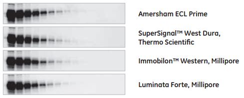 Fig 6. Amersham ECL Prime exhibits similar sensitivity as the competitor
reagents in Western blotting detection of ERK 1/2. The Millipore reagents
were slightly less sensitivity and also reached saturation at the highest
concentrations (10 to 5 μg), decreasing the dynamic range of these reagents. |
Figure 7 shows Amersham ECL Prime in comparison
with Clarity™ Western ECL Substrate from Bio-Rad.Samples: Two-fold dilution series of HeLa cell lysate starting at 2.5 μg
Primary antibody dilution: 1:3000
Secondary antibody dilution: 1:30 000
Detection: Amersham Imager 600, 3 min exposure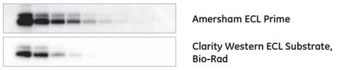 Fig 7. Amersham ECL Prime exhibits higher sensitivity and signal intensity
compared with Clarity Western ECL Substrate in Western blotting
detection of ERK 1/2. |
Amersham ECL
Figure 5 shows a side-by-side comparison of
Amersham ECL reagent with other chemiluminescence
detection reagents in the same sensitivity range.Samples: Two-fold dilution series of HeLa cell lysate starting at 5 μg
Primary antibody dilution: 1:1000
Secondary antibody dilution: 1:10 000
Detection: Amersham Imager 600, 1 min exposure |
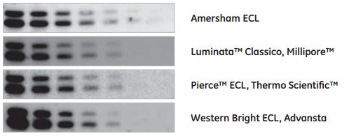
Fig 5. Amersham ECL reagent exhibits similar sensitivity as the competitor
reagents in Western blotting detection of ERK 1/2.
|

 생명과학
Cytiva


